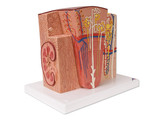
The 3B MICROanatomy™ Kidney is an extremely detailed model which shows the morphologic/functional units of the kidney. The kidney structures are greatly magnified. Six model zones illustrate the following fine-tissue structures of the human kidney that serve in the production of urine:
Longitudinal section of a kidney
Section of renal cortex and renal medulla of the kidney
Wedge-shaped section of a kidney lobe with a diagrammatic depiction of three nephrons with Henle’s loops of different lengths and diagrammatic depiction of the vascular supply
Diagrammatic illustration of a kidney nephron with a short Henle’s loop and didactic/diagrammatic illustration of the vascular supply
Diagrammatic illustration of an opened kidney renal corpuscle with nephron and light-microscopic transverse sections of the proximal, attenuated and distal segments of a renal tubule
Diagrammatic/didactic illustration of an opened kidney renal corpuscle
3B MICROanatomy™ Kidney mounted on a base.
Properties
- 309013
- K13

